Anatomy Of Musckes Sndctendons / Four muscles and their attached tendons make up the rotator cuff.. The shoulder is not a single joint, but a complex arrangement of bones, ligaments, muscles, and tendons that is better called the shoulder girdle. They are the continuations of muscles and allow them to connect to bones. They are associated with muscles discussed in the section above (see above). Related posts of diagram of shoulder muscles and tendons muscle anatomy human foot. Specific muscles are associated with specific movements of parts of the anatomy.
Lesson on the anatomy of the forearm: Tendons attach muscle to bone. It also helps you raise and rotate your arm. A tendon connects the muscle to the bone. The shoulder is not a single joint, but a complex arrangement of bones, ligaments, muscles, and tendons that is better called the shoulder girdle.
Achilles Tendon And Superficial Calf Muscle Anatomy Uptodate from www.uptodate.com It is separated from the first layer of muscles by the lateral plantar vessels and nerve. This video also provides you with a. The upper arm is located between the shoulder joint and elbow joint. In this lesson, we look at the muscle. Four muscles and their attached tendons make up the rotator cuff. The fleshy, thick part of the muscle is called its belly. Muscle anatomy gluteus 12 photos of the muscle anatomy gluteus gluteus muscle anatomy ct, gluteus muscle anatomy mri, human muscle anatomy gluteus maximus, muscle anatomy gluteus, muscle anatomy of gluteal, human muscles, gluteus muscle anatomy ct, gluteus muscle anatomy mri, human muscle anatomy gluteus maximus. The shoulder is not a single joint, but a complex arrangement of bones, ligaments, muscles, and tendons that is better called the shoulder girdle.
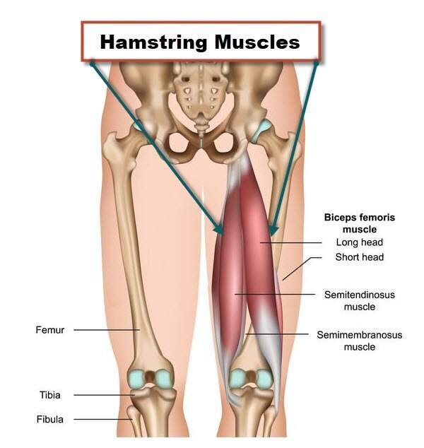
The leg anatomy includes the quads, hams, glutes, hip flexors, adductors & abductors.
The upper arm is located between the shoulder joint and elbow joint. The muscles of the hand can be broken down into three main regions: This video also provides you with a. Major muscles of the ankle. It attaches to the tendons of flexor digitorum longus. *the origin, insertion, and belly.* a muscle's origin is where a tendon attaches it to the *less* movable bone. For example, the sternocleidomastoid muscle (neck area) assists with movement of the head, while the psoas major muscle (low back area) is associated with flexion of the thigh. Related posts of diagram of shoulder muscles and tendons muscle anatomy human foot. Tendons and ligaments are bands of connective tissue that help stabilize the body and allow movement. They are associated with muscles discussed in the section above (see above). Each of them aids in a specific motion of your shoulder. On the other hand, the insertion is where a tendon attaches that muscle to the *more* movable bone. For that reason, and because of the dexterity of the shoulder joint itself, the musculature of the shoulder is complex, ranging from massive prime mover muscles to finer stabilizer and fixator muscles.
Tendons connect the knee bones to the leg muscles that move the knee. These muscles allow the ankle to bend downward and outward. Related posts of muscles and tendons of the leg muscle anatomy gluteus. There are numerous tendons around the knee that also help to stabilize the knee. Each of them aids in a specific motion of your shoulder.
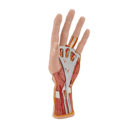
Life Size Hand Model With Muscles Tendons Ligaments Nerves Arteries 3 Part 3b Smart Anatomy 1000349 3b Scientific M18 Hand And Arm Skeleton Models Human Bone from www.3bscientific.com In this lesson, we look at the muscle. It also helps you raise and rotate your arm. This video also provides you with a. Tendons are the reason a muscle can move the bones in our body when muscles contract. Four muscles and their attached tendons make up the rotator cuff. Tendons and ligaments are bands of connective tissue that help stabilize the body and allow movement. In humans, the foot is one of the most complex structures in the body. The wrist links the hand to the forearm.
Tendons connect the knee bones to the leg muscles that move the knee.
The quadriceps muscles provide strength and power with knee extension (straightening). The shoulder is not a single joint, but a complex arrangement of bones, ligaments, muscles, and tendons that is better called the shoulder girdle. Learn about the muscles, tendons, bones, and ligaments that comprise the knee joint anatomy. Skeletal muscles are attached to bones by tendons and can be as long as 30 cm, although they are usually 2 to 3 cm in length. In this lesson, we look at the muscle. Every skeletal muscle has three main parts: Extensor carpi radialis brevis extensor carpi radialis longus The thenar (lateral or thumb side of the palm), hypothenar (medial or little finger side of the palm) and intermediate (middle of the hand) muscles. Tendons vary in size and are somewhat elastic and attach bones to muscles. Muscles and tendons of upper leg. The thenar muscles, which form the bulge of muscles evident at the base of the thumb, are essential to the hand's flexibility. In humans, the foot is one of the most complex structures in the body. The fleshy, thick part of the muscle is called its belly.
Related posts of diagram of shoulder muscles and tendons muscle anatomy human foot. Most of the muscles which act on the wrist joint are situated within the forearm, with only the tendon crossing the joint and inserting on the hand. The foot is a part of vertebrate anatomy which serves the purpose of supporting the animal's weight and allowing for locomotion on land. This is lesson 1 on the anatomy of the forearm. These muscles allow the ankle to bend downward and outward.
Anatomy Of Knee from marvel-b1-cdn.bc0a.com These muscles allow the ankle to bend downward and outward. Anatomy ankle anatomy ankle + ligament + tendon the foot anatomy human ankle anatomy 3d leg muscle lower leg anatomy leg articulation peroneal ankle muscles foot. The thenar muscles, which form the bulge of muscles evident at the base of the thumb, are essential to the hand's flexibility. Originates from the medial and lateral plantar surface of the calcaneus. In this lesson, we look at the muscle. The smaller bone that runs alongside the tibia (fibula) and the kneecap (patella) are the other bones that make the knee joint. Take this specially designed quiz to test your knowledge about the hand and wrist. The muscles you probably know the best are your.
The wrist links the hand to the forearm.
When the muscle contracts, the tendons are pulled, and the bone is moved. The muscles you probably know the best are your. Lesson on the anatomy of the forearm: The thenar (lateral or thumb side of the palm), hypothenar (medial or little finger side of the palm) and intermediate (middle of the hand) muscles. Take this specially designed quiz to test your knowledge about the hand and wrist. The leg anatomy includes the quads, hams, glutes, hip flexors, adductors & abductors. Tendons and ligaments are bands of connective tissue that help stabilize the body and allow movement. Similar to ligaments, they are made of collagen and can withstand increased tension. Über 7 millionen englischsprachige bücher. Muscle anatomy human foot 12 photos of the muscle anatomy human foot muscle anatomy human foot, muscle anatomy of the human foot, human muscles, muscle anatomy human foot, muscle anatomy of the human foot The muscles of the hand can be broken down into three main regions: The quadratus plantae muscle is located superior to the flexor digitorum longus tendons. The upper arm is located between the shoulder joint and elbow joint.


0 Komentar